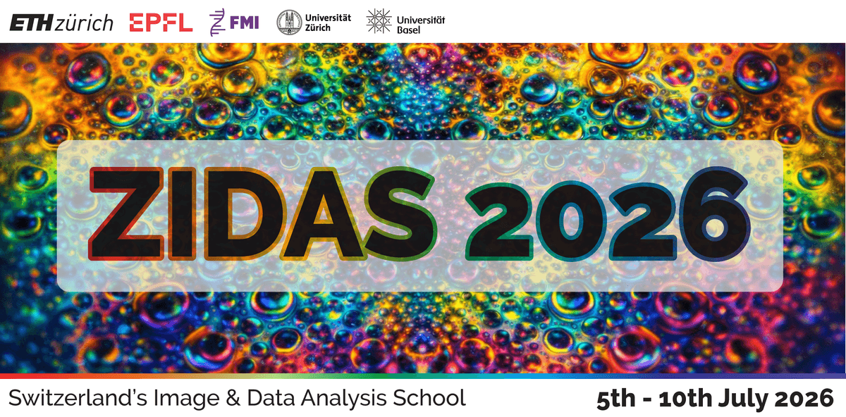
• ZIDAS main page

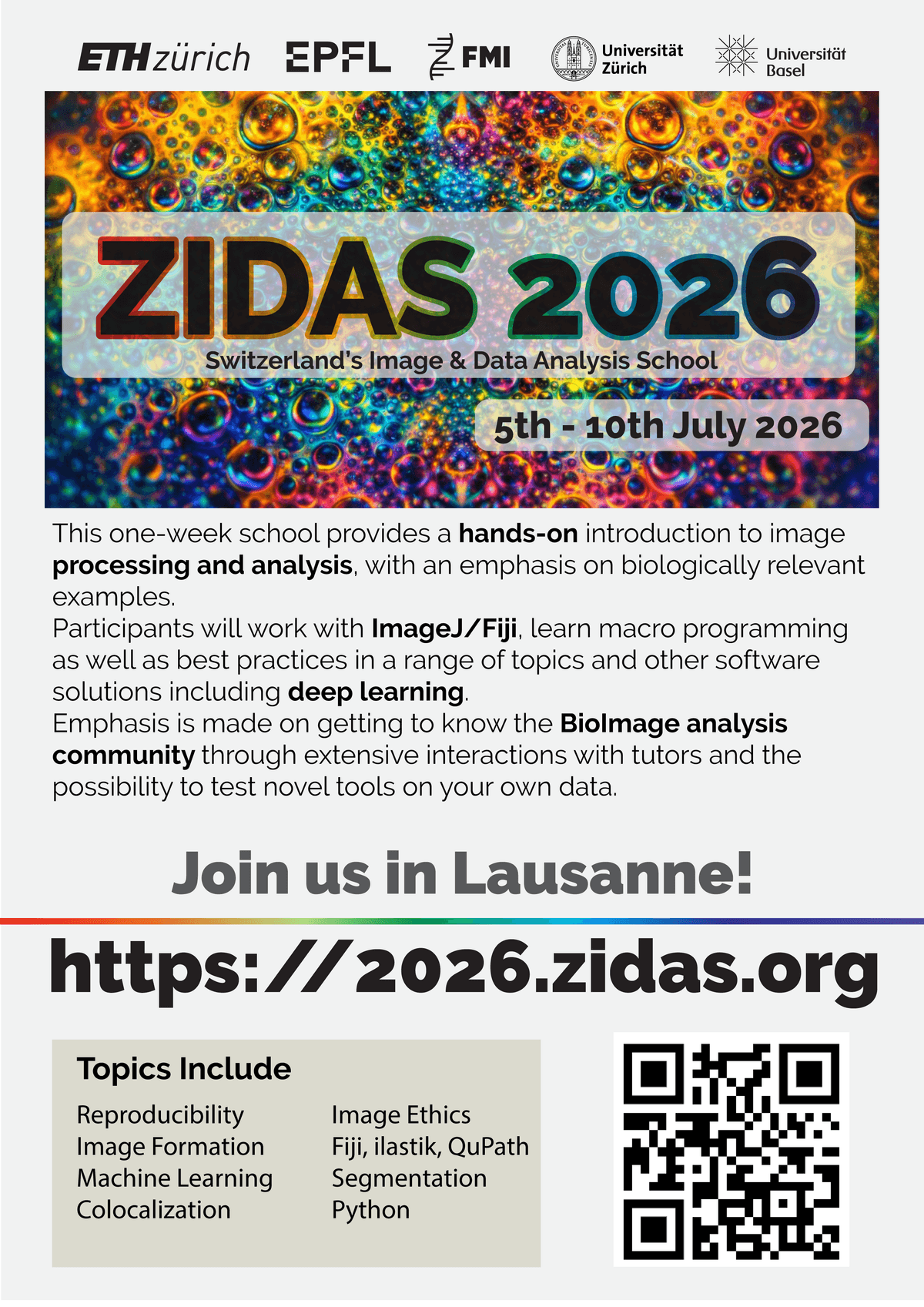
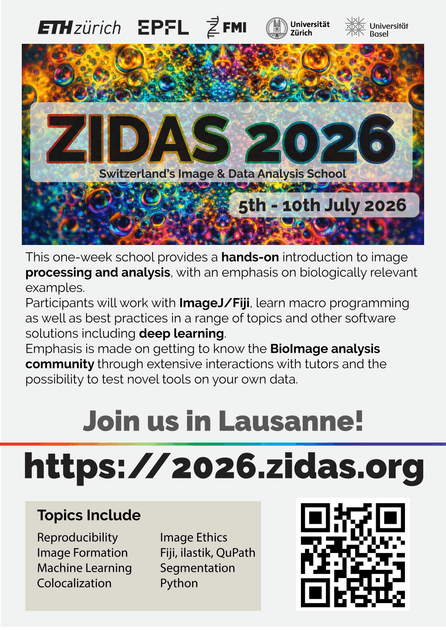
    Time: 5th - 10th July 2026

    Place: EPFL, Lausanne, Switzerland (in person)

  • Application is closed!

    Check back at a later date for information about future years.

  • About the School

    This one-week school provides a hands-on introduction to image processing and analysis, with an emphasis on biologically relevant examples.

    Is this school for you?

    • Are you a life-science researcher with a pressing need to quantify your light-microscopy images?
    • Are you uncertain about how to: Best calculate co-localisation, do deconvolution, automate the counting of cells, track objects over time, handle massive amounts of image data, record your image-analysis workflows in a reproducible manner?
    • If you answered yes to some of the above, then this school is for you!

    Motivation

    Digital images of high quality and quantity are now the norm in biomedical sciences. Ten to twenty years ago, when many current professors trained as students or post-docs, this was not yet the case as most microscopes were, at best, equipped with low-resolution digital cameras, celluloid-film (analogue) cameras, or no camera at all.


    This rapid change is rarely reflected in the curricula of life-science university departments and they offer few, if any, courses in image processing and analysis. Understandably, courses in image-analysis (computer-vision) in the computer-science departments tend to have different aims, work on different image-data, and pre-suppose literacy in at least one programming language, rendering them all but irrelevant for the life-scientist working in the laboratory.


    To bridge this gap, between what the life-scientist needs and what courses she/he is normally offered, we have created this school for image analysis. No former experience with programming is assumed, nor will much indeed be needed; only a strong desire to learn what can be done and how to do it is required.

    Structure of the week

    You will be working actively with image-analysis software every day -- this is an interactive hands-on school, not a passive lecture series. Short introductions are followed by guided workflows that we step through together. You can (and should) ask questions at any time throughout.


    There will be a single invited lecture every day, alternating between scientists using image-analysis as an integral part of their biomedical research and researchers developing new image-analysis algorithms and software.


    Also, we encourage you to bring your own data. During the training you will have a chance to explore various tools and concepts of image analysis and if they will be compatible, test them on your data, with support from the trainers.

    Financial support

    We are providing financial support to allow more people to apply despite financial constraints. We are going to provide: fee-waiver, travel support (up to 500CHF), and free accommodation for the period of training. Since we have only a limited number of scholarships, please apply for them only when your presence critically depends on receiving one.

    Factoids

    1. We will accept 25 participants --- this number is kept low to facilitate effective tutoring.
    2. You will need to bring your laptop and data.
    3. Participation is only possible for the entire week.
    4. The fee covers the attendance to the course and some snacks and drinks throughout the day. You need to cover travel, lunches & accommodation yourself.
    5. 3 x fee-waivers & travel grants & free accommodation are available and granted based on excellence & justified economical situation.
    6. The workshop starts on Sunday, with some team building activities. We plan dinner on Wednesday (covered in the participation fee!) and some casual networking activities during the week.
    7. PhD students can earn two ECTS credits from the school (we provide documents, upon approval of your local doctoral school).
  • Get pdf version

    Poster

    Please help us promote the event!

  • (Tentative) Program

    Expect some changes as we are working on the 2026 program right now...

  • Speakers

    Scientists using or developing image analysis methods in their research

    Section image

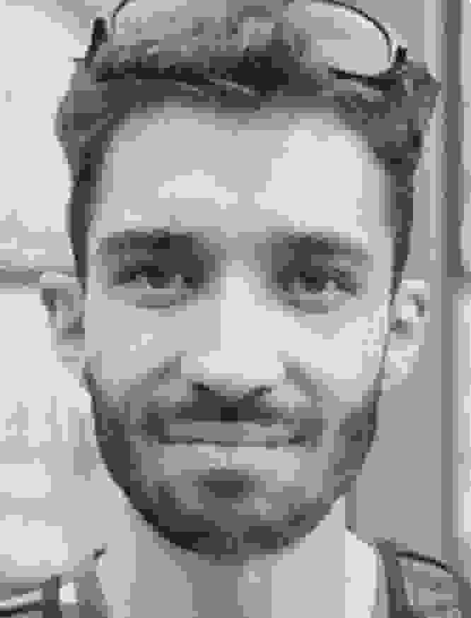
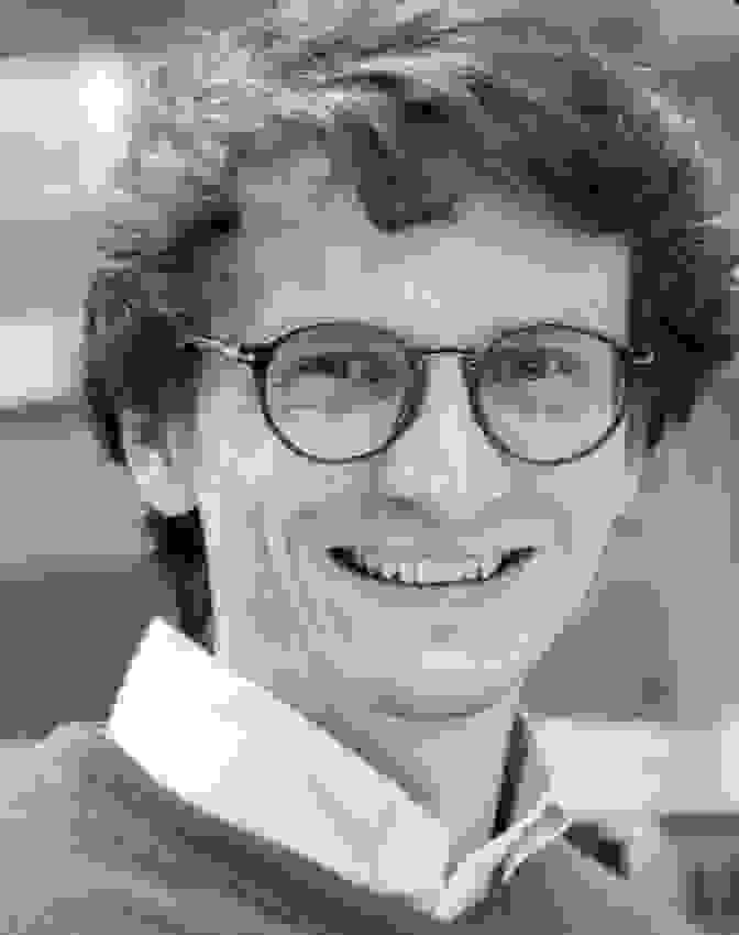
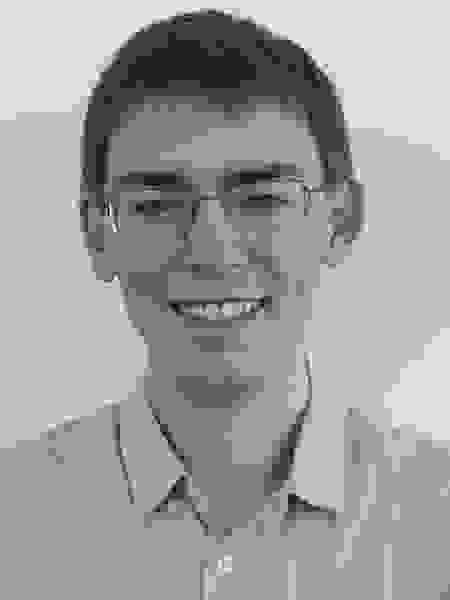
    Mackenzie Mathis is Associate Professor at EPFL and Bertarelli Foundation Chair of Integrative Neuroscience at the Brain Mind Institute. Her lab studies the neural circuits and computations behind adaptive behavior, with a focus on motor learning and control. She combines neuroscience, mouse models, quantitative behavior analysis, and deep learning, and also serves as a member of EPFL’s Open Science Strategic Committee.

    Section image

    Feyza Nur Arslan is a scientist in the Oates Lab at EPFL, working at the interface of cell and developmental biology, biophysics, and synthetic biology. Her research focuses on the mechanics of cell to cell adhesion and tissue development, including E cadherin mediated contacts and zebrafish embryogenesis. She brings experience from IST Austria, Bilkent University, EMBL, MIT, and Max Planck, with a strong interest in quantitative biology and science advocacy.

    Section image

    David Sholto

    Independent Consultant

    Sholto David is a microbiologist and science integrity investigator known for identifying errors, manipulated images, and unreliable data in published biomedical research. His work uses close visual inspection and AI tools such as ImageTwin, with findings shared through platforms including PubPeer and For Better Science. In 2024, TIME named him to the TIME100 Health list for his role in exposing flawed studies and pushing for stronger standards in scientific publishing.

  • Connect With Us

    Something unclear? Let us know!

  • Support and Endorsements

    This school was created by ScopeM-IDA staff, co-organized with BIOP, University of Basel, and FMI for Biomedical Research staff, powered by SwissBIAS trainers, and supported by EXCITE and Imaging@EPFL.

    Section image
    Section image
    Section image
    Section image
    Section image
    Section image
    Section image
    Section image
    Section image
    Section image
    Section image
  • Sponsors

    A great thank you to our wonderful sponsors for supporting open bioimage data analysis.

    Nikon
    ZEISS